
Mazabraud syndrome – case report and long term follow up


Authors:
- Ivar Vacula
Angiological Outpatient Clinic, Trnava - Roman Totkovič
Orthopedic Surgery Department, Hospital Košice-Šaca - Pavel Babál
Department of pathology, Faculty of Medicine, Comenius University, Bratislava
Case report
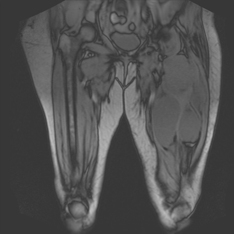
In January 2010 a 59 year old woman was presented to our angiological lab with the “edema” of her left thigh lasting for several weeks. Objectively, there were huge palpable tumors. Ultrasound imaging proved multiple encapsuled, hypoechogenic, non homogeneous soft tissue tumors in left quadriceps muscle, right great gluteal muscle and in pelvic region on the left side, almost surrounding common and external iliac artery and vein. No pathological lymphatic nodes were detected. An MRI and biopsy were performed to exclude sarcoma, with the result confirming intramusculary myxoma. Past history of our patient is remarkable. In 1983 she suffered a traumatic fracture of left proximal femur which was treated conservatively. In 1989, a solitary myxoma was removed from her right gluteal muscle. In 1998, a fibrous dysplasia of both iliac bones, left femur and tibia were diagnosed. Until 2010, no new soft tissue tumors were revealed.
Because of the progression of tumors from 2010, we performed angiography aimed for possible therapeutic embolization, but we failed to find pathologic vascularization. In 03/2010, based on biopsy results and imaging – the final diagnosis of Mazabraud syndrome (MS) was constituted. Our patient underwent two consecutive successful operations to decrease the amount of tumorous masses in her left thigh and right gluteal region (Picture1a,b). She is still followed, mostly using ultrasound imaging. Our experiencies suggest, that probably the tumors, which look hypoechogenic and homogenous have higher potential to grow compared to those, where there is more fibrous tissue – hyperechogenic septa (Picture 2a,b). CT or MRI is indicated approximately once in 2 years (Picture 3).
Discussion
MS is a very rare condition, firstly described by Henschen in 1926.1 The real number of reported cases remains unknown, a report from 2015 has been counted as 93th.2 As far as we know, our report is the first description from Slovakia. The prognosis of the disease is very good. It has no impact on life expectancy and usually a very limited impact on the quality of life. More serious complaints and danger is described in relation to fibrous bone dysplasia (deformities, fractures or pain and approximately 1 % risk of malignancy) compared to benign myxomas3,4. Our patient suffers from secondary lymphedema (Picture 4) of the left leg and pain in the left groin, mostly due to progression of the myxomas. We are reconsidering the next operation or alternative way how to diminish or desintegrate the tumors.
Conclusion
MS is a rare benign condition. The appropriate treatment of osteopenia or osteoporosis together with sporadic surgical removal of myxomas are well accepted and will be sufficient for most of these patients. Regular ultrasound and optional MRI or CT should be used for appropriate timing of interventions. We hope, that also our description will contribute to the general awareness of this uncommon disease.
Literature
- Henschen F. Fall von ostitis fibrosa mit multiplen tumoren in der umgebenden muskulatur. Verh Dtsch Ges Pathol 1926; 21:93–97.
- Shuiting Fu, Zhuowei Tian, Chenping Zhang and Yue He. Monostotic fibrous dysplasia and solitary intramuscular myxoma of the head and neck: A unique presentation of Mazabraud’s syndrome and a literature review. Oncology Letters 10: 3087-3094, 2015.
- Kransdorf MJ, Moser RP Jr, Gilkey FW. Fibrous dysplasia. Radiographics 1990;10:519-37.
- Samper Wamba JD, Fernandez Bermudez MJ, Dominguez TL, Pascua LR. Polyostotic fibrous dysplasia associated with intramuscular myxomas: Mazabraud syndrome. Indian J Radiol Imaging 2015;25:280-3.
 / Picture 1 (AFC AFS before)")


